Human skull inferior view mandible removed. Components and divisions of.
Hip Joint Illustrations Image Radiopaedia Org
A Structural Classification of Synovial Joints Label the types of synovial joints.
. Structural features ligaments and associated tendons of the shoulder joint Art-labeling Activity. The right elbow joint medial view. The fibrous connection between a tooth and its sockets is functionally and structurally classified as which of the following.
Pancreas larynx aorta Art-labeling Activity. Bone Markings Part 1. There is a printable worksheet available for download here so.
Adenine subunit Start codon mRNA strand A 498 g sample of aniline C6H5NH2 molar mass 9313 gmol was combusted in a bomb. It bears our bodys weight and the force of the strong muscles of the hip and leg. 35 Label The Parts Of The Lymphatic System - Labels Information List.
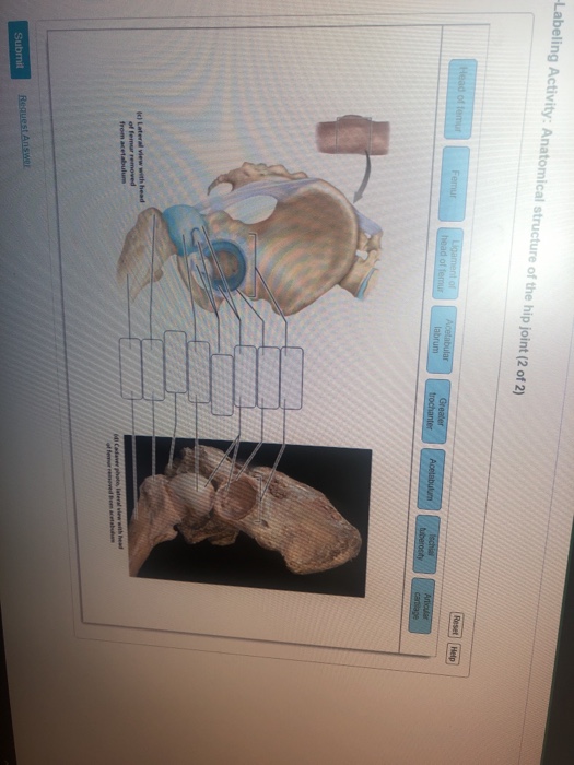
Start studying Mastering AP Chapter 7 -The Skeleton Art-labeling Activity. The Knee Joint Drag the correct label to the appropriate structure of the knee joint. Yet the hip joint is also one of our most flexible joints and allows a greater range of motion than all other joints in the body except for the shoulder.
Pivot joint Saddle sellaris joint Ball-and-socket joint Ellipsoid condylar joint Hinge joint Gliding planar joint Help Reset Eversion Protraction Inversion Depression Dorsiflexion Lateral. To allow for flexion the __________ unlocks the knee joint. The structure of a long bone humerus of arm Figure 59.
Compared to the glenohumeral shoulder joint however this joint sacrifices mobility for stability as it is designed for weight bearing. The hip joint is a multiaxial joint and permits a wide range of motion. Learn vocabulary terms and more with flashcards games and other study tools.
In ad-dition the bile salts facilitate the action of the en-zymes and help with the absorption of lipids through. A first carpometacarpal and metacarpophalangeal joints b glenohumeral joint c ankle joint d proximal radio-ulnar joint D. Structural features ligaments and associated tendons of the shoulder joint Art-labeling Activity.
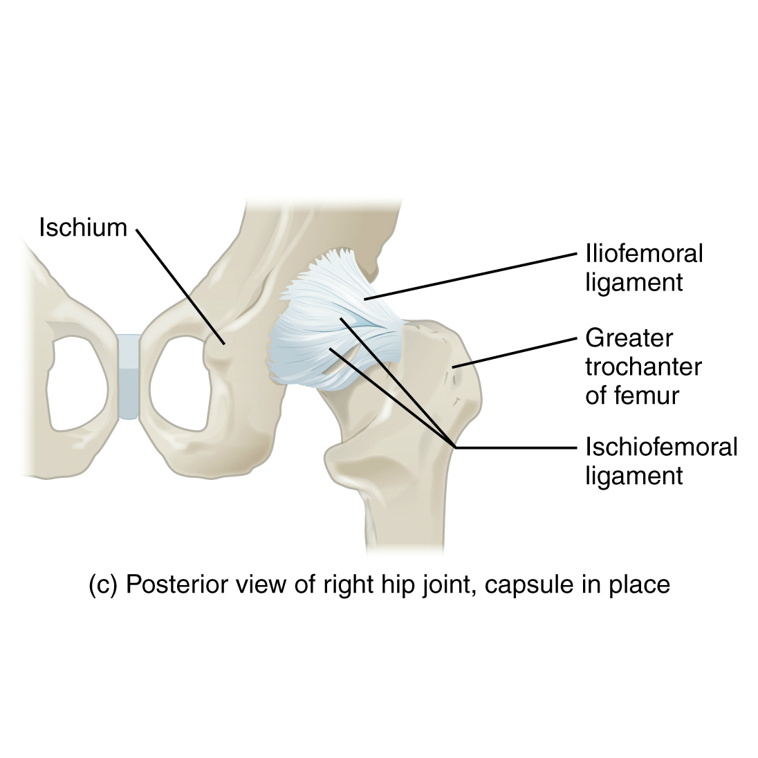
Popliteus The tibialis posterior muscle originates at which three locations. Muscles that move the hand and fingers anterior view middle layer Art-labeling Activity. The hip joint is a ball-and-socket synovial joint formed between the os coxa hip bone and the femur.
Types of special joint movements PICTURE Reading Quiz - Chapter 8 Question 5 At which joint do pronation and supination occur. Synovial Joints Identify synovial joints. JOHN Terms in this set 29 Drag the labels onto the diagram to identify structural features associated with the extrinsic muscles that move the foot and toes.
The Process of Translation Art-labeling Activity. Human skull superior view top of cranium removed Figure 511. Human skull lateral view.
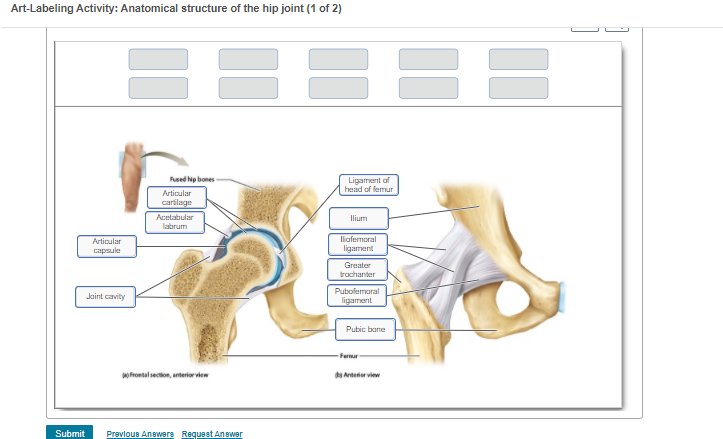
2 The body is divided into two major portions. Anatomy and Physiology questions and answers Art-labeling Activity The Right Hip Joint Acetabulum Transverse acrtabalar ligament Ischiateral the removed Greatertrochantar Ligament of the Publunan gent Acabularium os Lester och An anterior view Adore Show additional mesto streng to the cu esc c G Search or type URL 7 2 3 5 4 7. Learn vocabulary terms and more with flashcards games and other study tools.
C It has two layers an inner layer and an outer layer. March 13 2022 Reading time. Flexion extension abduction adduction external rotation internal rotation and circumduction.
Smooth muscle tissue in the walls of the calyces and renal pelvis contracts to help propel urine toward the ureter. ANATOMY 2220 MASTERING HW 2 OSU. Pivot joint Saddle sellaris joint Ball-and-socket joint Ellipsoid condylar joint Hinge joint Gliding planar joint Help Reset Eversion Protraction Inversion Depression Dorsiflexion Lateral flexion Plantar flexion Retraction Opposition Elevation.
Angular Movements of the Joints. It consists of two layers. Reset Help Fibula Lateral meniscus Anterior cruciate ligament Ligaments That Stabilize the Knee Joint Tbial collateral ligament Articular cartilage Fibular collateral ligament Patellar surface.
Activity art pancreas the. CISES 16-6 B On This Picture Draw And Label The F. A generalized nephron and collecting system.
The axial region includes the head neck and trunk. Chapter Test - Chapter 8 Question 12. Reset Help Fibula Lateral meniscus Anterior cruciate ligament Ligaments That Stabilize the Knee Joint Tbial collateral ligament Articular cartilage Fibular collateral ligament Patellar surface Tibia Patelar igament cut Posterior cruciate ligament Medial meniscus.
The right knee joint anterior view superficial layer Art-labeling Activity. Use the art-labeling activities to quiz yourself on key anatomical structures in this chapter. Written By jacksonesenwein71831 April 25 2022 Add Comment Edit.
Bone Markings on the Right Femur Learning Goal. Start studying Art-labeling Activity.
Art Labeling Activity Anatomical Structure Of The Chegg Com
Solved Ex 09 Hw Art Labelling Activity The Bones And Chegg Com

Solved Art Labeling Activity Anatomical Structure Of The Chegg Com
Solved Ex 09 Hw Art Labelling Activity The Bones And Chegg Com
Solved Labeling Activity Anatomical Structure Of The Hip Chegg Com

Hip Joint Radiology Reference Article Radiopaedia Org
2
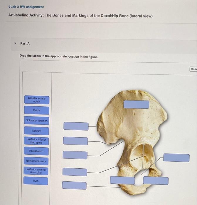
Solved Lab 3 Hw Assignment Art Labeling Activity Regions Chegg Com
0 comments
Post a Comment